Imaging Center
The Neuroscience Imaging Center is located on the third floor of the Link building. It provides services in light and electron microscopy to faculty, staff and graduate students at the University of Tennessee, as well as the Memphis research community and private institutions nationwide.
Operating on a cost recovery basis, it offers use of equipment and technical services in the areas listed below.
JEOL 2000EX with a high resolution digital camera and monitor
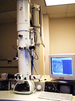
Normally running at 60kV, this electron microscope can be used at higher kV's to accommodate the need for thicker sections.
High-resolution digital images can be routinely captured in many different formats, and stored on CD's or DVD's. Images can be viewed between 60x and 1000x in the Low Magnification mode, and between 600x and 1,000,000x in the regular Magnification mode.
Location and Scheduling
Located on the 3rd floor of the Link building in room 311. Please visit the web calendar to schedule equipment use. New users, please contact Esther Marquez Wilkins at 901.448.5976 or emarque2@uthsc.edu for instructions and signup.
Zeiss 710 with 34-Channel Spectral Detection, 6 Laser Lines, and 6144 x 6144 Resolution
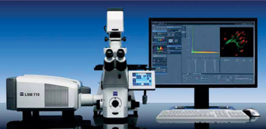
The Zeiss 710 features 34-channel spectral detection (10 nm resolution), 7 laser lines (405 nm, 458 nm, 488 nm, 514 nm, 561 nm, 594 nm and 633 nm) and 6144 x 6144 resolution. This system has live imaging capability, and advanced imaging methods such as FRET (Forster resonance energy transfer), FRAP (fluorescence recovery after photobleaching), tiling, stitching.
The system is attached to a Zeiss Axio Observer inverted microscope.
Location and Scheduling
Located on the 3rd floor of the Link building in room 314. Please visit the web calendar to schedule equipment use. New users, please contact Esther Marquez Wilkins at 901.448.5976 or emarque2@uthsc.edu for instructions and signup.
Microbright Field Inc. NeuroLucida
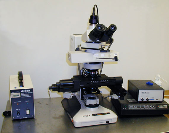
The Neurolucida software allows 3D reconstruction of neurons, serial reconstruction of brain sections, and precise cell plotting in real-time.
Neurolucida360 software allows for 3D reconstruction of neurons obtained from Z-stacks from fluorescent confocal microscopes.
Stereology
Stereo Investigator is advanced scientific software for unbiased design-based stereology. Stereo Investigator can analyze data from multiple modalities: using live images from digital or video cameras or stored image sets from confocal microscopes, electron microscopes, and scanning tomographic sources. It also features sophisticated tools for anatomical mapping, which can be used to delineate regions of interest for stereology probes, to map cell distributions, to prepare anatomical maps for publication, and to perform detailed morphometric analyses.
Location and Scheduling
Located on the 3rd floor of the Link building in room 310. Please visit the web calendar to schedule equipment use. New users, please contact Esther Marquez Wilkins at 901.448.5976 or emarque2@uthsc.edu for instructions and signup.
Conventional Light and Fluorescence Microscopy
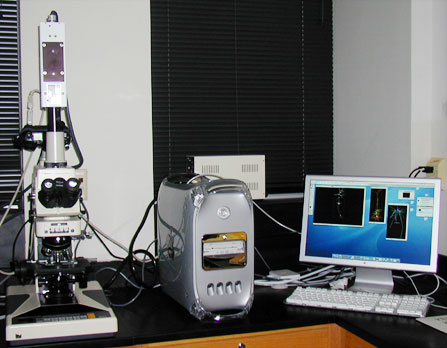
We have a Nikon Microphot FX microscope capable of light and dark field imaging as well as epifluorescence. The high resolution Kodak DCS 460 color digital camera is mounted on the microscope and can be moved to a camera stand with a light box. Using a PowerPC G4, images are viewed on an Apple 20" Cinema Display monitor.
Reichert Jung UltraCut E Microtome
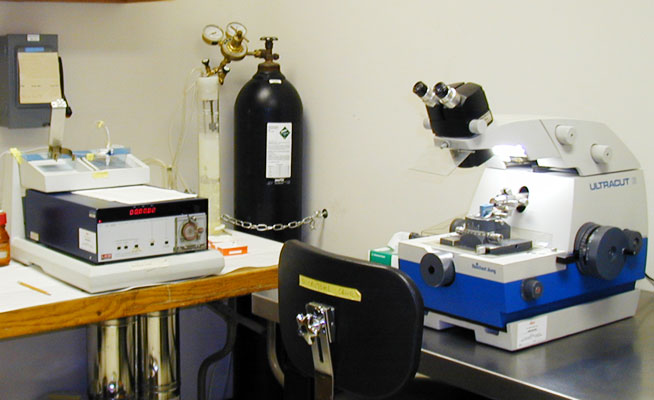
Tissue can be embedded is Spurr's resin, Epon, or Epon/Araldite blends, as well as LRWhite for immunocytochemistry. This lab has experience processing many types of tissue, tissue slices, tissue cultures, and pellets. A variety of embedment methods are available, such as Beem or gelatin capsules, flat molds, and flat embedment.
Thin Sectioning
Thin sections, ranging from 50nm to 200nm in thickness, are routinely cut on the Reichert Jung UltraCut E microtome. Many types of grids are available to accommodate the investigators needs.
Semi-Thin Sectioning
Semi-thin sections, also called thick sections, are readily cut on the Reichert Jung UltraCut E microtome, using glass knives. Counter-staining with Toluidine Blue is available.
Location and Scheduling
Located on the 3rd floor of the Link building. Please visit the web calendar to schedule equipment use. New users, please contact Esther Marquez Wilkins at 901.448.5976 or by emarque2@uthsc.edu for instructions and signup.
Negative Staining
Negative staining can be done in the lab using uranyl acetate or phosphotungstic acid and viewed immediately on the electron microscope.
Liquid Nitrogen Availability
Two large tanks of liquid nitrogen are on hand for those investigators who need it on the occasional basis.